• Blog
  • About
  • Contact
Jenny Jozz

Therapy And Severs Disease

5/19/2015

 
Overview

Sever's disease or Calcaneal apophysitis is a condition that affects children between the ages of 10 and 13 years. It is characterized by pain in one or both heels with walking. During this phase of life, growth of the bone is taking place at a faster rate than the tendons. Hence there is a relative shortening of the heel-cord compared to the leg bones. As a result, the tension the heel-cord applies to the heel bone at its insertion is very great. Moreover, the heel cord is attached to a portion of the calcaneus (heel bone) that is still immature, consisting of a mixture of bone and growing cartilage, called the calcaneal apophysis, which is prone to injury. Compounding to this is the fact that all these changes are happening in a very active child, prone to overuse. The end result is therefore an overuse syndrome of injury and inflammation at the heel where the heel cord (Achilles Tendonitis) inserts into the heel bone (Calcaneal apophysitis).

Causes

This condition is more common in boys than girls. It generally presents between the ages of 9-14 and peaks between ages 10-12 years. This injury can reoccur up until the age of 17, when the growth plate of the calcaneous generally closes. These types of injuries will commonly occur during periods of rapid growth. Sever?s Disease occurs more frequently in child with flat feet, but all children with flat feet will not get Sever?s.

Symptoms

In Sever?s disease, heel pain can be in one or both heels. It usually starts after a child begins a new sports season or a new sport. Your child may walk with a limp. The pain may increase when he or she stands on tiptoe. Your child?s heel may hurt if you squeeze both sides toward the very back. This is called the squeeze test. Your provider may also find that your child?s heel tendons have become tight.

Diagnosis

Sever's disease is diagnosed based on a doctor?s physical examination of the lower leg, ankle, and foot. If the diagnosis is in question, the doctor may order X-rays or an MRI to determine if there are other injuries that may be causing the heel pain.

Non Surgical Treatment

Treatment is primarily supportive, with rest, pain management, and activity modification. Activity modifications include the addition of low-impact activities. Gel heel cups are sold over the counter and can be used intermittently to help reduce shock in the heel, as well as take tension off of the tight Achilles?s tendon complex. Proper stretching and strengthening activities should be preformed routinely even during periods of no pain. A large study showed that approximately 85% of children affected by Sever?s disease return to full activity within a two-month time period after starting treatment.

Prevention

Properly stretching to maintain flexibility is effective for preventing Sever's disease. Stretches should target the calves, heel cords, and hamstrings. Your child should do the appropriate stretches two or three times a day, holding the stretch for about 20 seconds each time. Ask your child's doctor for specific exercise instructions. Generally, doctors advise stretching both legs, even if the pain is confined to one heel. It's also helpful to strengthen the shin muscles by having your youngster pull his toes in with a rubber exercise band or a piece of tubing and then stretch them forward. Assist your child in doing 15 repetitions of this exercise, three times a day. Having your child wear shoes with good shock absorbers and avoid running on hard surfaces as much as possible should also help prevent the condition.

Pain In The Arch What Are The Causes ?

4/16/2015

 
Overview

Plantar fasciitis is an inflammation of a thick, fibrous ligament in the arch of the foot called the plantar fascia. The plantar fascia attaches into the heel bone and fans out toward the ball of the foot, attaching into the base of the toes. If this ligament is stretched excessively it will become inflamed and begin to cause pain. In severe instances the ligament can rupture resulting in immediate severe pain. If the ligament ruptures the pain is so great that the patient can not place weight on the foot. Should this happen, the foot should be elevated and an ice pack applied. An appointment with your foot doctor should be made at your earliest convenience.

Pain In Arch

Causes

The most common cause of arch pain is plantar fasciitis. Plantar fasciitis is inflammation of the fibrous band of tissue that connects the heel to the toes. Sufferers of plantar fasciitis typically feel pain early in the morning when they first get out of bed. Another common cause of arch pain is fallen arches or flat feet. Fallen arches result in the sole of the foot becoming flat so that the entire sole of the foot touches the ground. Fallen arches can lead to pain in the ankles, knees, feet, and even in the legs. While fallen arches are often inherited, for some it develops as a result of wear and tear on your feet. Wearing shoes without proper metatarsal support, or arch support, can overstretch the tendon that supports the arch causing the arch to flatten out.

Symptoms

Typically, the sufferer of plantar fasciitis experiences pain upon rising after sleep, particularly the first step out of bed. Such pain is tightly localized at the bony landmark on the anterior medial tubercle of the calcaneus. In some cases, pain may prevent the athlete from walking in a normal heel-toe gait, causing an irregular walk as means of compensation. Less common areas of pain include the forefoot, Achilles tendon, or subtalar joint. After a brief period of walking, the pain usually subsides, but returns again either with vigorous activity or prolonged standing or walking. On the field, an altered gait or abnormal stride pattern, along with pain during running or jumping activities are tell-tale signs of plantar fasciitis and should be given prompt attention. Further indications of the injury include poor dorsiflexion (lifting the forefoot off the ground) due to a shortened gastroc complex, (muscles of the calf). Crouching in a full squat position with the sole of the foot flat on the ground can be used as a test, as pain will preclude it for the athlete suffering from plantar fasciitis, causing an elevation of the heel due to tension in the gastroc complex.

Diagnosis

The diagnosis of high arch (cavus) foot deformity or Charcot Marie Tooth disease can be made by an orthopedic surgeon in the office. Evaluation includes a thorough history and physical examination as well as imaging studies such as X-rays. The orthopedic surgeon will look at the overall shape, flexibility, and strength of a patient?s foot and ankle to help determine the best treatment. Nerve tests may occasionally need to be performed to help confirm the diagnosis.

Non Surgical Treatment

An orthotic arch support, specially molded to fit your foot, may be part of your treatment. These supports can be particularly helpful if you have flat feet or high arches. You can tell if that is what is needed when short-term taping decreases your heel pain.

Arch Pain

Surgical Treatment

Surgery may be necessary in situations where the symptoms are likely to get worse over time, or when pain and instability cannot be corrected with external orthopedic devices. There are many types of surgical procedures, including cavus foot reconstruction, which can be performed to correct the foot and the ankle and restore function and muscle balance.

Prevention

To prevent arch pain, it is important to build up slowly to your exercise routine while wearing arch supports inside training shoes. By undertaking these simple measures you can prevent the discomfort of arch pain which can otherwise linger for many months. While you allow the foot to recover, it will help to undertake low impact exercises (such as swimming or water aerobics).

Achilles Tendon Rupture The Facts

4/12/2015

 
Overview Achilles Tendonitis A rupture of the Achilles tendon is one of the more common types of tendon ruptures. As the largest tendon in the human body, the Achilles tendon is very important to one?s balance and stability. Patients who suffer an Achilles tendon rupture experience severe pain and often require Achilles tendon surgery to repair the damage. There are also less invasive methods of Achilles tendon treatment including, icing, anti-inflammatory medications, rehabilitation, taping or bracing, sports massages and more. Achilles tendon recovery is dependent on the health of the patient, and the severity of the injury. Causes An Achilles tendon injury might be caused by several factors. Overuse. Stepping up your level of physical activity too quickly. Wearing high heels, which increases the stress on the tendon. Problems with the feet, an Achilles tendon injury can result from flat feet, also known as fallen arches or overpronation. In this condition, the impact of a step causes the arch of your foot to collapse, stretching the muscles and tendons. Muscles or tendons in the leg that are too tight. Achilles tendon injuries are common in people who participate in the following sports. Running. Gymnastics. Dance. Football. Baseball. Softball. Basketball. Tennis. Volleyball. You are more likely to tear an Achilles tendon when you start moving suddenly. For instance, a sprinter might get one at the start of a race. The abrupt tensing of the muscle can be too much for the tendon to handle. Men older than age 30 are particularly prone to Achilles tendon injuries. Symptoms Typically patients present with sudden onset of pain and swelling in the achilles region, often accompanied by a audible snap during forceful dorsiflexion of the foot. A classic example is that of an unfit 'weekend warrior' playing squash. If complete a defect may be felt and the patient will have only minimal plantar flexion against resistance. Diagnosis During the physical exam, your doctor will inspect your lower leg for tenderness and swelling. In many cases, doctors can feel a gap in your tendon if a complete rupture has occurred. The doctor may also ask you to kneel on a chair or lie on your stomach with your feet hanging over the end of the exam table. He or she may then squeeze your calf muscle to see if your foot will automatically flex. If it doesn't, you probably have ruptured your Achilles tendon. If there's a question about the extent of your Achilles tendon injury, whether it's completely or only partially ruptured, your doctor may order a magnetic resonance imaging (MRI) scan. This painless procedure uses radio waves and a strong magnetic field to create a computerized image of the tissues of your body. Non Surgical Treatment This condition should be diagnosed and treated as soon as possible, because prompt treatment probably improves recovery. You may need to be referred urgently to see a doctor in an orthopaedic department or accident and emergency department. Meanwhile, if a ruptured Achilles tendon is suspected, you should not put any weight on that foot, so do not walk on it at all.Treatment options for an Achilles tendon rupture include surgical and non-surgical approaches. The decision of whether to proceed with surgery or non-surgical treatment is based on the severity of the rupture and the patient?s health status and activity level. Non-surgical treatment, which is generally associated with a higher rate of re-rupture, is selected for minor ruptures, less active patients, and those with medical conditions that prevent them from undergoing surgery. Non-surgical treatment involves use of a cast, walking boot, or brace to restrict motion and allow the torn tendon to heal. Achilles Tendinitis 
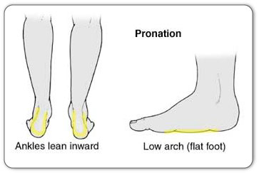
Surgical Treatment Surgery will involve stitching the two ends of the tendon together, before placing the leg in a cast or brace. The advantage of having an operation is the reduced chance of the rupture reoccurring, however it will involve the risks associated with any surgical procedure, such as infection. Prevention To reduce your chance of developing Achilles tendon problems, follow the following tips. Stretch and strengthen calf muscles. Stretch your calf to the point at which you feel a noticeable pull but not pain. Don't bounce during a stretch. Calf-strengthening exercises can also help the muscle and tendon absorb more force and prevent injury. Vary your exercises. Alternate high-impact sports, such as running, with low-impact sports, such as walking, biking or swimming. Avoid activities that place excessive stress on your Achilles tendons, such as hill running and jumping activities. Choose running surfaces carefully. Avoid or limit running on hard or slippery surfaces. Dress properly for cold-weather training and wear well-fitting athletic shoes with proper cushioning in the heels. Increase training intensity slowly. Achilles tendon injuries commonly occur after abruptly increasing training intensity. Increase the distance, duration and frequency of your training by no more than 10 percent each week.

Pes Planus Causes, Conditions And Treatment Solution

4/2/2015

 
Overview

Flat Feet

Fallen arches, the medical term for flat feet, are simply feet that do not arch in the middle. Such feet lay flat with the whole of the sole on the ground. All children are born with flat feet, but as they grow the feet normally develop an arch so that by the age of ten almost all children have arched feet. Occasionally a child will not develop properly arched feet. This may be due to a bone malformation present at birth, or abnormalities in ligaments within the feet or legs that cause the child's weight to be unevenly distributed.


Causes

Genetic predisposition. Faulty foot mechanics, e.g. excessive pronation. Abnormal bony architecture. Laxity of ligaments. Neuro-muscular disease. Trauma to the leg muscles or major tendons. Inflammatory diseases of the joints e.g. Rheumatoid arthritis. Surgical procedures on the leg and ankle. Limb length inequality. Tight Achilles tendon.


Symptoms

Many people have flat feet and notice no problems and require no treatment. But others may experience the following symptoms, Feet tire easily, painful or achy feet, especially in the areas of the arches and heels, the inside bottom of your feet become swollen, foot movement, such as standing on your toes, is difficult, back and leg pain, If you notice any of these symptoms, it's time for a trip to the doctor.


Diagnosis

Many medical professionals can diagnose a flat foot by examining the patient standing or just looking at them. On going up onto tip toe the deformity will correct when this is a flexible flat foot in a child with lax joints. Such correction is not seen in the adult with a rigid flat foot. An easy and traditional home diagnosis is the "wet footprint" test, performed by wetting the feet in water and then standing on a smooth, level surface such as smooth concrete or thin cardboard or heavy paper. Usually, the more the sole of the foot that makes contact (leaves a footprint), the flatter the foot. In more extreme cases, known as a kinked flatfoot, the entire inner edge of the footprint may actually bulge outward, where in a normal to high arch this part of the sole of the foot does not make contact with the ground at all.


Non Surgical Treatment

Foot orthotic devices can stabilize some of the causes of flat feet, optimize muscle function and reduce the occurrence of painful symptoms. Generally, a custom-made semi-rigid functional posted orthotic is effective for flat feet. These devices are prescribed based on a thorough biomechanical examination by a qualified chiropodist/podiatrist. Over-the-counter arch supports may be helpful for mild cases, but often prove ineffective to relieve symptoms associated with flatfoot. Wear shoes with a good fit in the arch. Keep active and fit to strengthen leg and foot musculature. Control body weight to decrease load on the feet. Avoid prolonged periods weight bearing with bare feet. Taping to support strained ligaments and joints. Physical therapy modalities such as ultrasound. Referral for surgical correction a severe symptomatic flatfoot.


Surgical Treatment

Adult Acquired Flat Foot

Surgery is typically offered as a last resort in people with significant pain that is resistant to other therapies. The treatment of a rigid flatfoot depends on its cause. Congenital vertical talus. Your doctor may suggest a trial of serial casting. The foot is placed in a cast and the cast is changed frequently to reposition the foot gradually. However, this generally has a low success rate. Most people ultimately need surgery to correct the problem. Tarsal coalition. Treatment depends on your age, extent of bone fusion and severity of symptoms. For milder cases, your doctor may recommend nonsurgical treatment with shoe inserts, wrapping of the foot with supportive straps or temporarily immobilizing the foot in a cast. For more severe cases, surgery is necessary to relieve pain and improve the flexibility of the foot. Lateral subtalar dislocation. The goal is to move the dislocated bone back into place as soon as possible. If there is no open wound, the doctor may push the bone back into proper alignment without making an incision. Anesthesia is usually given before this treatment. Once this is accomplished, a short leg cast must be worn for about four weeks to help stabilize the joint permanently. About 15% to 20% of people with lateral subtalar dislocation must be treated with surgery to reposition the dislocated bone.


Prevention

Orthotic inserts, either prescribed or bought over the counter, can help keep the arches fixed into position, but always wear them as although they support, they don?t strengthen, which is why some experts reccomend avoiding them. Gait analysis at a run specialist can help to diagnose overpronation and flat feet. Most brands produce shoes that will give support and help to limit the negative effects of a poor gait on the rest of the body. Barefoot exercises, such as standing on a towel and making fists with the toes, can help to strengthen the arches. Start easy and build up the reps to avoid cramping. Short barefoot running sessions can help take pressure off the arches by using the natural elasticity of the foot?s tendons to take impact and build strength to help prevent flat feet. These should be done on grass for only a few minutes at a time.

What Can Cause Pain In The Arches ?

3/28/2015

 
Overview

Plantar fasciitis is a common, painful foot condition. Patients, and sometimes doctors often confuse the terms plantar fasciitis and heel spurs. Plantar fasciitis refers to the syndrome of inflammation of the band of tissue that runs from the heel along the arch of the foot; a heel spur is a hook of bone that can form on the heel bone (calcaneus). About 70% of patients with plantar fasciitis have been noted to have a heel spur that can be seen on x-ray.

Foot Arch Pain

Causes

A person with flat feet has greater load placed on the posterior tibial tendon which is the main tendon unit supporting up the arch of the foot. Throughout life, aging leads to decreased strength of muscles, tendons and ligaments. The blood supply diminishes to tendons with aging as arteries narrow. Heavier, obese patients have more weight on the arch and have greater narrowing of arteries due to atherosclerosis. In some people, the posterior tibial tendon finally gives out or tears. This is not a sudden event in most cases. Rather, it is a slow, gradual stretching followed by inflammation and degeneration of the tendon. Once the posterior tibial tendon stretches, the ligaments of the arch stretch and tear. The bones of the arch then move out of position with body weight pressing down from above. The foot rotates inward at the ankle in a movement called pronation. The arch appears collapsed, and the heel bone is tilted to the inside. The deformity can progress until the foot literally dislocates outward from under the ankle joint.

Symptoms

Go to a podiatrist at the first sign of symptoms. Besides pain on the bottom of the foot, additional symptoms may include burning sensation in arch, difficulty standing on tiptoes, inflammation, more pain after sleeping or resting, redness, heat, localized pain in the ball of the foot, sharp or shooting pain in the toes, pain that increases when toes are flexed, tingling or numbness in the toes, aching, pain that increases when walking barefoot, pain that increases when walking on hard surfaces, pain the increases when standing (putting weight on your feet) or moving around and decreases when immobile, skin Lesions, it?s important to get a proper diagnosis and treatment plan. Let?s go over the possible causes of the pain.

Diagnosis

A patient is asked to step with full body weight on the symptomatic foot, keeping the unaffected foot off the ground. The patient is then instructed to "raise up on the tip toes" of the affected foot. If the posterior tibial tendon has been attenuated or ruptured, the patient will be unable to lift the heel off the floor and rise onto the toes. In less severe cases, the patient will be able to rise on the toes, but the heel will not be noted to invert as it normally does when we rise onto the toes. X-rays can be helpful but are not diagnostic of the adult acquired flatfoot. Both feet, the symptomatic and asymptomatic - will demonstrate a flatfoot deformity on x-ray. Careful observation may show a greater severity of deformity on the affected side.

Non Surgical Treatment

There is considerable debate about the best treatment option for plantar fasciitis. Some authors suggest all of the 'mainstream' methods of treatment don't actually help at all and can actually make the symptoms worse! However, on the whole, there are several of the most commonly cited treatment options for plantar fasciitis and these are generally accepted throughout the medical community. I would recommend giving these options a try if you haven't already. Rest. This is mainly applicable to the sports people as rest is possible treatment. (For those who cannot rest e.g. people who work on their feet - skip to the other treatment options below). Rest until it is not painful. This is made more difficult as people need to use their feet to perform daily activities but certainly stop sporting activities that are likely to be putting the fascia under excessive stress. Perform Self Micro-Massage (you can watch this video by clicking the link or scrolling further down the page as it's embedded on this lens!) This massage technique is used to break down fibrous tissue and also to stimulate blood flow to the area, both of which encourage healing and reduce pain. There is also a potentially soothing effect on nerve endings which will contribute to pain relief. Ice Therapy. Particularly useful after spending periods on your feet to reduce the inflammation. Wrap some ice or a bag of frozen peas in a towel and hold against the foot for up to 10 minutes. Repeat until symptoms have resolved. Heat Therapy. Heat therapy can be used (not after activity) to improve blood flow to the area to encourage healing. A heat pack of hot water bottle can be used. 10 minutes is ideal. Careful not to burn yourself! A good taping technique. By taping the foot in a certain way you can limit the movement in the foot and prevent the fascia from over-stretching and gives it a chance to rest and heal. Click on the link for more information on taping techniques. Weight Management. If you are over-weight, any weight you can loose will help to ease the burden on your sore feet and plantar fascia. Orthotic devices (often mis-spelled orthodic) are special insoles that can be used to limit over-pronation (discussed earlier) and control foot function. By preventing the arches flattening excessively, the plantar fascia is not over-stretched to the same extent and this should help with the symptoms and encourage healing. Stretching the calf muscles (again, click this link or scroll to the bottom of the page to watch the embedded video) can help to lengthen these muscles and the Achilles tendon - a risk factor for plantar fasciitis. Stretching of the plantar fascia itself is also encouraged, particularly before getting up the morning (night splints can be used for this effect) and after periods of rest. This can be achieved by placing a towel or band under the ball of the foot and gently pulling upwards until a stretch is felt. Hold for about 15-20 seconds then rest briefly. Repeat 2-3 times. As you can see there are many different treatment options available. Try incorporating some of these in to your daily routine and see what works for you. Regardless of the method the main aim is to prevent the fascia from over-stretching! Medical professionals such as a Podiatrist may decide to make custom orthotics or try ultra-sound therapy. It is likely that anti-inflammatory medications will also be recommended. If you have tried the treatment options and your symptoms persist I'd recommend going to see a medical professional for further advice.

Arch Pain

Surgical Treatment

The soft tissue surgeries usually would include a lengthening of the Achilles tendon, releasing of the plantar fascia as well as tendon transfers. These procedures are usually done in conjunction with bony procedures such as calcaneal osteotomies (to lower the heel bone and get it more under the leg itself), as well as metatarsal osteotomies. These procedures usually involve either cutting or fusion of the bones, and placement of fixation devices to allow the bones to heal. Healing time is usually at least 6-8 weeks and usually the patient must be non-weight bearing during the healing process. These types of surgical corrections are usually reserved for the more difficult, painful and deformed feet. They can require more surgeries down the line. These procedures are usually the last resort after all other modes of treatment have been exhausted (except in children where it is usually best to treat the deformity early). There are many different degrees of high arched feet and these procedures should be left for the more extreme cases. These cases usually require a very high degree of surgical skill and should only be done by those who frequently perform these types of cases.

Prevention

Stretch and strengthen important muscles in your feet, ankles and legs in order to guard against future strain. Make sure to acquire suitable arch supports and inserts if necessary, and that your shoes are shock absorbent and in good condition. Wearing tattered shoes provides no protection, and runners should replace their footwear before exceeding 500 miles of usage. Athletes new to arch supports should gradually build their training routine, allowing their feet to become accustomed to a new stance.

Stretching Exercises

Stretching your calf and Achilles tendon may also help as a tight Achilles can make your foot overpronate. To stretch your calf and Achilles tendon, step forwards with your left leg and bend it, with your right leg straight and both feet pointing forwards. Push your right heel into the ground while keeping your right leg straight; you should feel the stretch at the back of your right leg, below the knee. Hold the stretch for 15 to 30 seconds and repeat with the opposite leg. Repeat the stretch two to four times on each leg, and repeat the overall exercise three to four times a day.

What's Adult Aquired Flat Foot ?

3/26/2015

 
Overview

Noticed that will your own foot is actually receiving flatter plus more painful? Carry out you've trouble walking or even performing exercise exercise without having leg and arch pain? Have you heard the term "fallen arches"? Almost All associated with this stuff make reference to an ailment known as posterior tibial tendon dysfunction. This particular is definitely an inflammation along with overuse syndrome regarding certainly one of the extended tendons in which pass from the leg across the inside in the ankle and also attaches to the inside arch in the foot. the posterior tibial tendon?s job is to help offer the arch and allow for more effective gait.

Flat Feet

Causes

Overuse with the posterior tibial tendon will be often the reason for PTTD. Within fact, the signs as well as symptoms typically occur after activities which involve the tendon, such as running, walking, hiking, or perhaps climbing stairs.

Symptoms

Pain and swelling behind the interior of one's ankle as well as together your current instep. An Individual may always be tender powering the actual inner ankle the spot exactly where the posterior tibial tendon programs along with from time and energy to time acquire burning, shooting, tingling or even stabbing pain like a results of inflammation with the nerve inside the particular tarsal tunnel. Trouble walking, the particular inability to walk extended mileage along with a generalised ache while strolling even brief distances. This kind of may most likely grow in order to be more pronounced after each day. Adjust inside foot shape, sometimes your tendon stretches out, this can be because of in order to weakening in the tendon and also ligaments. Whenever this occurs, the actual arch inside your foot flattens along with a flatfoot deformity occurs, presenting the alteration of foot shape. Inability in order to tip-toe, the method of diagnosing Posterior Tibial Tendon Dysfunction is actually trouble as well as inability to be able to ?heel rise? (stand on your current own toes on one foot). The tibialis posterior tendon enables you to carry out this manoeuvre effectively. you could also experience pain upon attempting to do a new heel rise.

Diagnosis

Posterior Tibial Tendon Dysfunction is clinically decided to have careful clinical observation with the patient?s gait (walking), selection of motion testing for your foot along with ankle joints, and diagnostic imaging. people together with flatfoot deformity walk with the heel angled outward, in addition referred in order to as over-pronation. Though it truly is typical for your arch in order to impact the bottom regarding shock absorption, individuals with PTTD come using an arch which completely collapses to the ground along with will not reform an arch throughout the entire gait period. after evaluating the particular ambulation pattern, the actual foot and ankle array of motion needs to be tested. Generally the particular affected foot may have decreased motion for the ankle joint and furthermore the hindfoot. Muscle Mass strength might also become weaker as well. An straightforward test to perform for PTTD may become the single heel raise in which usually the patient is actually asked to raise up around the ball regarding their effected foot. A New normal foot sort could lift up around the toes with out pain and additionally the heel will invert slightly when the person offers completely raised your heel up during the test. Throughout early phases involving PTTD the individual may become able to lift up the actual heel but the heel is not really going to invert. An elongated or even torn posterior tibial tendon, which in turn is actually a mid for you to late finding of PTTD, will prohibit the actual patient via totally rising up around the heel and can cause intense pain for the arch. Lastly diagnostic imaging, although used alone can't diagnose PTTD, can easily provide further details with an accurate diagnosis of flatfoot deformity. Xrays with the foot can easily present the particular practitioner important angular relationships with the hindfoot as well as forefoot which in turn aid diagnose flatfoot deformity. The Majority Of with the time, an MRI is not required to diagnose PTTD nevertheless is truly a instrument that should always be considered throughout advanced cases of flatfoot deformity. In your event that any partial tear of the posterior tibial tendon is regarding concern, then an MRI could show your anatomic place with the tear and additionally the extensiveness of the injury.

Non surgical Treatment

Initial remedy relies about the level of deformity and flexibility with initial presentation. Conservative remedy includes orthotics or perhaps ankle foot orthoses (AFO) to offer the posterior tibial tendon (PT) as well as the longitudinal arch, anti-inflammatories to assist decrease pain and inflammation, exercise modification which might consist of immobilization of the foot and physical therapy to help strengthen along with rehabilitate the particular tendon.

Adult Acquired Flat Foot

Surgical Treatment

Surgical correction would depend on the severity of signs along with symptoms and in addition the stage involving deformity. The Particular goals involving surgery tend to be to generate a far more functional and steady foot. There are multiple treatments available towards the surgeon and it usually takes several to be able to correct any flatfoot deformity. Generally surgical treatment starts off with removal of inflammatory tissue along with repair in the posterior tibial tendon. A New tendon transfer will be performed when the posterior tibial muscle is actually weak or possibly the tendon will be badly damaged. Your the majority of widely used tendon is the flexor digitorum longus tendon. This kind of tendon flexes or even moves your lesser toes downward. Your flexor digitorum longus tendon is utilized thanks to become able to its close proximity for the posterior tibial tendon and because you will find minimal unwanted effects with its loss. The Actual remainder with the tendon is actually sutured for the flexor hallucis longus tendon that flexes the important toe to ensure that small function will be loss.

Pain In The Foot's Arch Cause And Effect

3/23/2015

 
Overview

Each foot contains 26 bones, which form two arches. The longitudinal arch runs the length of the foot, and the transverse arch runs the width. The bones of the arch are primarily held together by the shape with which they fit with each other and by fibrous tissues known as ligaments that serve to hold the bones to each other. The muscles of the foot, along with a tough, sinewy tissue known as the plantar fascia, provide secondary support to the foot. There are also fat pads in the foot to help with weight-bearing and absorbing impact. Arch pain can occur whenever something goes wrong with the function or interaction of any of these structures.

Foot Arch Pain


Causes

Foot cramps are caused by muscles suddenly spasming uncontrollably. They most commonly cause foot arch pain but can occur anywhere in the foot and lower leg. Usually, they only last a few seconds but in more extreme cases they can continue longer. Often, there is no obvious reason why people suffer from foot cramps, but possible causes include diet, muscle tightness and weakness, dehydration, reduced circulation and fatigue. Sometimes, it can be a sign of an underlying medical condition so if the problem keeps recurring, do consult your doctor. Some of the best ways to reduce the incidence of foot arch pain from cramps include doing exercises, using heat, drinking plenty of water, using toe stretchers and ensuring you are wearing good footwear.


Symptoms

Symptoms of plantar fasciitis may occur anywhere along the arch, but it is most common near its attachment to the heel bone. Symptoms of plantar fasciitis vary, but the classic symptom is pain after rest--when you first get out of bed in the morning, or when you get up after sitting down for a while during the day. This is known as "post-static dyskinesia." The pain usually diminishes after a few minutes of walking, sometimes even disappearing, but the pain is commonly felt again the longer you're on the foot. Fasciitis can be aggravated by shoes that lack appropriate support, especially in the arch area, and by the chronic irritation of long-periods of standing, especially on concrete, and by being overweight. Other factors which influence this condition are gender (females get this more than men), age (30s to 50s are most common), and those with flatter-than-normal feet. It doesn't help that fascia doesn't heal particularly quickly. This is because it has relatively poor circulation, which is why it's white in colour.


Diagnosis

A patient is asked to step with full body weight on the symptomatic foot, keeping the unaffected foot off the ground. The patient is then instructed to "raise up on the tip toes" of the affected foot. If the posterior tibial tendon has been attenuated or ruptured, the patient will be unable to lift the heel off the floor and rise onto the toes. In less severe cases, the patient will be able to rise on the toes, but the heel will not be noted to invert as it normally does when we rise onto the toes. X-rays can be helpful but are not diagnostic of the adult acquired flatfoot. Both feet, the symptomatic and asymptomatic - will demonstrate a flatfoot deformity on x-ray. Careful observation may show a greater severity of deformity on the affected side.


Non Surgical Treatment

Arch pain can be treated with orthotics, inserts that have proper arch support to relieve the strain on the plantar fascia, mild stretching and anti-inflammatory medications. Orthotics will relieve most of the strain put onto the plantar fascia by supporting the band from underneath when pressure is applied. Tape can also be used in conjunction with orthotics to restrict movement and support the plantar fascia. Stretching should be used along with orthotics and continued long after the symptoms of arch pain are gone to prevent it from occurring again.

Pain In Arch


Surgical Treatment

Surgical advances have dramatically improved the ability to alleviate the pain and decreased function that millions of Americans experience due to flat feet. Nevertheless, many patients and even some physicians remain unaware of the new procedures, which are best performed by a foot and ankle specialist who has the applicable training and experience.


Prevention

Stretch and strengthen important muscles in your feet, ankles and legs in order to guard against future strain. Make sure to acquire suitable arch supports and inserts if necessary, and that your shoes are shock absorbent and in good condition. Wearing tattered shoes provides no protection, and runners should replace their footwear before exceeding 500 miles of usage. Athletes new to arch supports should gradually build their training routine, allowing their feet to become accustomed to a new stance.


Stretching Exercises

Strengthening exercises. Below are two simple strength exercises to help condition the muscles, tendons and joints around the foot and ankle. Plantar Rolling. Place a small tin can or tennis ball under the arch of the affected foot. Slowly move the foot back and forth allowing the tin can or tennis ball to roll around under the arch. This activity will help to stretch, strengthen and massage the affected area. Toe Walking. Stand upright in bare feet and rise up onto the toes and front of the foot. Balance in this position and walk forward in slow, small steps. Maintain an upright, balanced posture, staying as high as possible with each step. Complete three sets of the exercise, with a short break in between sets, for a total of 20 meters.

Arch Pain Causes Signs And Symptoms And Treatment Procedures

3/22/2015

 
Overview

One of those often-painful soft tissue that attaches to heel spurs at the bottom of the foot is called "plantar fascia". Fascia, located throughout the body, is a fibrous connective tissue similar to a ligament. You can see fascia when you handle meat. It is the white, connective tissue separating layers of meat or attaching to bones. The "plantar" fascia in our bodies is that fascia which is seen on the bottom (or plantar portion) of the foot, extending from the heel bone to the ball of the foot. Compared to other fascia around the body, plantar fascia is very thick and very strong. It has to be strong because of the tremendous amount of force it must endure when you walk, run or jump. But while the plantar fascia is a strong structure, it can still get injured, most commonly when it is stretched beyond its normal length over long periods of time. When plantar fascia is injured, the condition is called "plantar fasciitis", which is usualy pronounced either "plan-tar fash-I-tis" or "plan-tar-fash-ee-I-tis." (Adding "-itis" to the end of a word means that structure is inflamed.) It is sometimes known more simply as 'fasciitis'. Plantar fasciitis is the most common type of arch pain.

Pain In Arch


Causes

Having low or no arches is normal for some people. In these cases, flat feet are usually inherited and the feet are fairly flexible. Occasionally, flat feet can be caused by an abnormality that develops in the womb, such as a problem with a joint or where two or more bones are fused together. This is known as tarsal coalition and results in the feet being flat and stiff. Flat feet that develop in later life can be caused by a condition that affects the joints, such as arthritis, or an injury to a muscle, tendon or joint in the foot.


Symptoms

Symptoms include pain which is often described as a burning pain radiating into the arch of the foot, heel and sometimes the toes. Pins and needles or numbness may be felt in the sole of the foot. Pain may be worse when running or when standing for long periods of time and often worse at night. The area under the medial malleolus on the inside of the ankle may be tender to touch.


Diagnosis

The adult acquired flatfoot, secondary to posterior tibial tendon dysfunction, is diagnosed in a number of ways with no single test proven to be totally reliable. The most accurate diagnosis is made by a skilled clinician utilizing observation and hands on evaluation of the foot and ankle. Observation of the foot in a walking examination is most reliable. The affected foot appears more pronated and deformed compared to the unaffected foot. Muscle testing will show a strength deficit. An easy test to perform in the office is the single foot raise.


Non Surgical Treatment

Treatment for a high arch foot or Charcot Marie Tooth disorder depends on the extent of deformity and the amount of disability experienced by the patient. Depending upon the symptoms, treatment may include. Changing the shoes. Special orthotic supports (devices that support, adjust, or accommodate the foot deformity). Cushioning pads. Foot and ankle braces or surgery.

Foot Arch Pain


Surgical Treatment

As with most surgeries, patients and physicians should consider the surgery only after other, less invasive treatments have proven unproductive. Indications for surgery include Pain. Inability to function. Failure to improve after a six-month course of specific, directed physical therapy. Failure to improve after using arch supports, orthotics, or ankle and foot bracing. Once patients are at that point, the good news is that the procedure has considerably better outcomes than more traditional flat foot surgery. In the past, surgeons would realign and fuse the three hind joints, which would cause patients to lose motion, leaving them with a significantly stiff hind foot, With these newer procedures, if the foot is still flexible, surgeons can realign it and usually restore a close-to-normal or functional range of motion in the joints.


Prevention

Arch pain occurs when the plantar fascia becomes worn down due to constant strain or excessive exercising. This may be caused by increasing your running or hiking mileage too fast, wearing inadequate footwear, lack of stretching, running on steep hills, standing on your feet for too long and abnormal anatomy such as flat foot. Stretching is an important exercise that should not be overlooked because the tightness or lack of tightness of the joints in the foot can also cause pain in the arch.

Achilles Tendinitis

3/20/2015

 
Overview
Achilles Tendonitis The Achilles tendon is a conjoined tendon composed of the gastrocnemius and soleus muscles with occasional contribution from the plantaris muscle, and it inserts on the calcaneal tuberosity. The plantaris muscle is absent in 6% to 8% of individuals. The Achilles tendon is approximately 15-cm long and is the largest and strongest tendon in the human body. The tendon spirals approximately 90 from its origin to its insertion and this twisting produces an area of stress approximately 2- to 5-cm proximal to its insertion. The tendon has no true synovial sheath; instead it is wrapped in a paratenon. The Achilles tendon experiences the highest loads of any tendon in the body, and bears tensile loads up to 10 times body weight during athletic activities. The tendon most commonly ruptures in a region 2- to 6-cm proximal to its insertion.
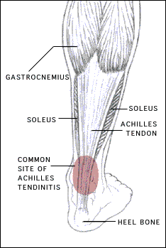
Causes
Causes of and contributors to Achilles tendon rupture include trauma (caused by injury, usually an acceleration injury such as pushing off or jumping up). Preceding tendon problems. Chronic Achilles tendonitis (can lead to small tears within the tendon, increasingly weakening it). Certain drug therapies/treatments. Drugs that have been linked to Achilles tendon rupture include. Fluoroquinolone antibiotics - after nearly 900 reports of tendon ruptures, tendonitis and other tendon disorders (most associated with the Achilles tendon) linked to Ciprofloxacin (Cipro) alone were collected in the U.S. Food and Drug Administration (FDA)s database, at least one public-interest group petitioned the FDA to recommend that a "Black Box Warning" be added to Cipro's packaging. Some researchers speculate this class of antibiotics is toxic to tendon fibers, and that in some cases may reduce their blood supply. Patients should at least be more aware of the potential for ruptures so that they can be switched to other antibiotics at the onset of early warning signals such as tendon pain.

Symptoms
Patients who suffer an acute rupture of the Achilles tendon often report hearing a popor snap. Patients usually have severe pain the back of the lower leg near the heel. This may or may not be accompanied by swelling. Additionally, because the function of the Achilles tendon is to enable plantarflexion (bending the foot downward), patients often have difficulty walking or standing up on their toes. With a complete rupture of the tendon, the foot will not move. In cases where the diagnosis is equivocal, your physician may order an MRI of the leg to diagnose a rupture of the Achilles tendon.

Diagnosis
An Achilles' tendon injury can be diagnosed by applying the Thompson Test (or Calf Squeeze Test) this is where the person who has suffered the injury lies on their front with their legs bent. Whoever is performing the test, usually a doctor, will then squeeze the calf muscle. If the tendon has not ruptured then the foot should point briefly away from the leg.

Non Surgical Treatment
Non-surgical treatment of Achilles tendon rupture is usually reserved for patients who are relatively sedentary or may be at higher risk for complications with surgical intervention (due to other associated medical problems). This involves a period of immobilization, followed by range of motion and strengthening exercises; unfortunately, it is associated with a higher risk of re-rupture of the tendon, and possibly a less optimal functional outcome. Achilles Tendonitis

Surgical Treatment
The goal of surgery is to realign the two ends of the ruptured tendon to allow healing. There are multiple techniques to accomplish this goal that will vary from surgeon to surgeon. Recovery from this injury is usually very successful with return to full function in approximately 6 months. Post operatively casting is required with the use of crutches or other means to remain non-weightbearing for 4-8 weeks. This is followed by a course of physical therapy. Partial rupture may or may not require surgical intervention depending on the extent of injury but cast immobilization is a common requirement.

Prevention
You can help to reduce your risk of an injury to your Achilles tendon by doing the following. When you start a new exercise regime, gradually increase the intensity and the length of time you spend being active. Warm up your muscles before you exercise and cool them down after you have finished. The benefit of stretching before or after exercise is unproven. However, it may help to stretch your calf muscles, which will help to lengthen your Achilles tendon, before you exercise. Wear appropriate and well-fitting shoes when you exercise.
Forward>>

    Author

    Write something about yourself. No need to be fancy, just an overview.

    Archives

    March 2015

    Categories

    All

    RSS Feed


Powered by Create your own unique website with customizable templates.